Research

Cell Analysis

Sed sed pharetra sem. Morbi fermentum mollis nisl ac sollicitudin. In ullamcorper mauris congue risus porta, non porta ante egestas. Class aptent taciti sociosqu ad litora torquent per conubia nostra, per inceptos himenaeos.

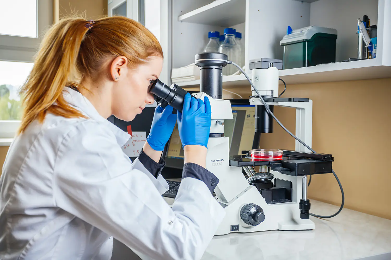
A female researcher in blue gloves observing cell samples under an Olympus microscope.

Unit Profile

Collaboration and services

Information regarding collaboration and applications will be expanded upon in the business module.

Staff

Pracownie

Histology

We provide services for the preparation and processing of tissue samples, the creation of histological sections, routine and specialized staining, and immunohistochemical and immunofluorescence analyses. We offer support in selecting and optimizing methods, as well as in interpreting results.

Preparation of Tissue Samples

Preparation of tissue sections using the paraffin and frozen sections methods, tailored to the purpose of the study. The process includes fixation (for paraffin sections), embedding of the material, cutting sections of appropriate thickness, staining, and mounting of microscopic slides. The prepared slides serve as material for histological, histochemical, immunohistochemical, and immunofluorescence studies, enabling the assessment of tissue structure and morphological changes.

Rutynowe analizy histologiczne

Wykonywanie standardowych barwień histologicznych umożliwiających ocenę budowy tkanek oraz identyfikację zmian patologicznych. W zależności od celu badania stosowane są m.in. barwienie hematoksyliną i eozyną (H+E), Luxol Fast Blue (LFB), trichrom Massona oraz reakcja PAS, pozwalające na ocenę struktury tkanek, włókien nerwowych, tkanki łącznej, glikogenu i innych składników komórkowych.

Oferta pracowni

Rutynowe analizy histologiczne:

  • Hematoksylina/Eozyna (H+E)
  • Barwienie Luxol Fast Blue (LFB)
  • Barwienie metodą Massona (Masson’s trichrome)
  • barwienie kwasem Schiffa (PAS)

Immunohistochemia i immunofluorescencja

Wykrywanie i lokalizacja wybranych białek oraz markerów komórkowych w tkankach z wykorzystaniem metod immunohistochemii (IHC) i immunofluorescencji (IF). Analiza umożliwia ocenę ekspresji białek, proliferacji komórek, procesów demielinizacji, nasilenia stanu zapalnego, włóknienia oraz integralności bariery krew–mózg. Badania znajdują zastosowanie w ocenie mechanizmów działania badanych substancji, biomateriałów i wyrobów medycznych oraz w badaniach przedklinicznych.

Oferta pracowni

Przykładowe markery:

  • Proliferacja: Ki-67, PCNA
  • Apoptoza: Caspase-3
  • Stan zapalny: Iba1, CD68, TNF-α, IL-1β
  • Demielinizacja: Iba1
  • Włóknienie: α-SMA, kolagen I, kolagen III, fibronektyna
  • Bariera krew–mózg: fibrynogen